


Get a personalized assessment of your operational efficiency and accelerate growth for your business.
The introduction of AI (Artificial Intelligence) in the medical imaging field is quite promising. It shows ingenious progress in the early detection and diagnosis of different diseases.
AI-based medical imaging software can offer viable solutions to the healthcare sector. It can efficiently handle and process the digital data produced during the scans and provide the most accurate results.
In this blog, we will look at how AI can help develop advanced medical imaging software and some impressive examples and use cases.
Let’s dive in.
How can AI Help in Medical Imaging?
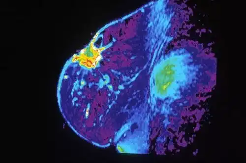
AI can improve the medical imaging process in hospitals in many ways. It is exciting to see the use of technology to reduce scan times and enhance patient care.

Photo Credit: vectorpouch / Freepik
The technology can help radiologists and other medical professionals improve productivity. Technologists reiterate that advanced medical imaging software is keeping the number of individuals in hospitals down, lowering the chance of transmission.
Such initiatives support the imaging community by making the best medical decisions. Let’s look at how AI can facilitate medical imaging. 1.
1. Higher Productivity with Automation
AI can automate a few parts of the radiology workflow. It can help analyze medical images faster than medical professionals, as it has better computational capabilities.
It can improve speed, efficiency, and accuracy, which can further lead to better care.
2. More Accurate Diagnosis
According to studies, AI can be more proficient than medical professionals and experts at diagnosing diseases such as cancer-based on medical images.
For example, scientists at Google have developed an AI that can facilitate in diagnosing breast cancer. The technology captures medical images via slides and uses deep learning algorithms to analyze cancerous cells.
The AI recorded 99% accuracy in cancer diagnosis, based on the slides corresponding to 38% of some doctors in the comparison group.
Also Read: The Essential Guide to IT Infrastructure Software Development
3. Computing Quantitative Data
AI can use quantitative data in multiple ways beyond the limits of human cognition.
It can predict if a patient will suffer from heart failure based on their rate of hospital visits and medical history.
The countless possibilities have led to a significant rise in AI-based imaging modality systems across the globe. The growth has fueled the rapid evolution in deep learning techniques and an increasing number of cross-industry partnerships.
Top Use Cases of AI in Medical Imaging
Let’s look at some of the real-life use cases of AI in medical imaging.
1. Screening Common Cancers
At heart, most software curators have the desire to improve the efficacy of clinical care, which includes AI.
Within cancer imaging, AI finds excellent utility in performing clinical tasks that include detection, characterization, and tumor monitoring.
- Detection: At this stage, AI-based detection tools can reduce observational oversights and errors of omission.
- Characterization: The task broadly captures the diagnosis, segmentation, and staging of tumors. It provides robust tumor descriptors that help to capture intra-tumor heterogeneity and variability.
- Monitoring: AI can play a significant role in monitoring changes in a tumor over time. Temporal tracking of tumor changes either in natural history or in response to treatment.
Medical imaging finds a significant spot in preventive screenings of cancers, such as lung cancer, breast cancer, and colon cancer.
For example, according to experts, medical imaging in AI plays a vital role in improving the early detection and characterization of lung cancer.
It can differentiate benign from malignant nodules. Early detection and improved accuracy can help improve patient outcomes and minimal overtreatment. The diseases detected at early stages are often curable.
Furthermore, AI can also enhance lung cancer staging and characterization and monitoring treatment response. It can use quantitative imaging features to categorize microcalcifications more accurately. In fact, the technology can potentially decrease the rate of unnecessary benign biopsies.
2. Identifying Risks for Cardiovascular Diseases
AI techniques such as machine learning (ML), cognitive computing, and deep learning (DL) have immense potential to change how cardiology and cardiovascular medicine are practiced, especially in cardiovascular imaging.
It can help measure the various structures of the heart and reveal an individual’s risk for cardiovascular diseases.
AI can identify problems that may need to be addressed through surgery or pharmacological management. Automating the detection of abnormalities in commonly-ordered imaging tests, such as chest x-rays, will not only expedite decision-making but also reduce diagnostic errors.
So, think for a moment, if a patient lands up in an emergency healthcare unit, complaining of shortness of breath, the chest radiograph as the first imaging study can be helpful.
It can be used as an initial screening tool for cardiomegaly, and medical professionals can use it as a marker for heart disease.
In fact, Zebra Medical Vision and Clalit Health Services presented a research project that allows the early identification of patients with cardiovascular disease using AI. Using existing computed tomography (CT) data, Zebra-Med’s AI algorithms allow Clalit find out patients who are at risk of cardiac events.
In the future, radiologists’ visual assessments that are sometimes erroneous can transcend to the efficient use of AI. For example, the identification of left atrial enlargement from chest x-rays could eliminate other cardiac or pulmonary problems.
Furthermore, it can help providers target appropriate treatments for patients. Subsequently, automated pulmonary artery flow quantification would save the interpreting physician time.
3. Accuracy in the Diagnosis of Neurological Diseases
Over the past decade, computed tomography (CT), positron emission tomography (PET), and magnetic resonance imaging (MRI) have revolutionized the study of the brain.
Experts estimate the day-to-day error rates and discrepancies in radiology are greater than 3%-5%. It is evident that novel methods and advanced medical imaging software can help physicians effectively analyze data.
The quality of medical data will increase, which can support better disease analysis and control.
Think for a moment—there is no cure for some degenerative neurological diseases, such as amyotrophic lateral sclerosis (ALS). In such cases, accurate diagnoses can help individuals understand likely outcomes and also prepare for long-term care.
Imaging studies are critical in identifying ALS and also differentiating ALS and primary lateral sclerosis (PLS). Radiologists play a crucial role in deciding if lesions are mimicking the structures of one of the diseases.
Medical professionals know that manual segmentation and quantitative susceptibility mapping (QSM) assessments of the motor cortex are difficult, necessary, and time-consuming.
ML techniques are now increasingly popular for addressing brain-related problems. Automating such procedures with ML could aid in the development of promising imaging biomarkers. Such novel initiatives can help reduce providers' workflow burdens.
4. Detecting Thoracic Complications
Pneumonia and pneumothorax can turn into a life-threatening emergency due to lung collapse and respiratory or circulatory distress. Delays in the detection and treating serious pneumothorax can result in severe harm to patients. In such cases, artificial algorithms can aid physicians.
Radiology images are popularly used to diagnose pneumonia and distinguish the condition from other lung conditions, such as bronchitis. However, radiologists aren’t always available to read images.
Even when radiologists are present, they may still have difficulty detecting pneumonia in cases where patients have pre-existing lung conditions, including cystic fibrosis or malignancies.
Here, an AI algorithm could assess x-rays and other images for evidence of opacities that indicate pneumonia. Subsequently, it can alert healthcare providers to the potential diagnoses and allow swifter treatment.
Furthermore, computer algorithms, backed with high-quality training data, could help detect pneumothorax on a chest X-ray with significant accuracy to help prioritize images for quick review by physicians.
Radiologists could potentially use algorithms as a tool to increase the speed with which a severe pneumothorax is detected, especially at times of lower staffing, when turnaround times are typically longer.
Let’s look at a good example that reiterates accuracy in the detection of thoracic complications and conditions. In a research study, algorithms could detect the majority (80%–84%) of images showing a moderate or large pneumothorax while correctly categorizing 90% or more of images without pneumothorax as negative.
So, the implementation of such algorithms can improve the speed and quality of care delivered across various healthcare settings. Plus, AI may also be able to help providers monitor patients over time.
Rapid detection and swift communication with treating medical professionals may result in faster treatment of pneumothorax and lessening the impacts of a severe medical problem.
Leading Examples of AI in Medical Imaging
Let’s look at some popular AI and machine vision technologies that are approved for clinical use.
1. QuantX (Quantitative Insights)
Paragon Biosciences and Qlarity Imaging have developed a way to harness the power of artificial intelligence to aid humans in catching cancer earlier and more accurately.
QuantX is the first-ever computer-aided breast cancer diagnosis system cleared by the FDA for use in radiology. It is a quantitative image analysis software device used to assist radiologists in assessing and characterizing breast abnormalities using MR image data.
The software has been helping radiologists interpret MRIs, noting the differences between cancerous and noncancerous breast lesions. As aptly stated by the CEO and chairman of Paragon Biosciences, radiology is the backbone of diagnosing many diseases, and the future is radiologists with technology.
Plus, QuantX may also be used as an image viewer of multi-modality digital images, including ultrasound and mammography. The software includes tools that allow users to measure and document images and output in a structured report.
2. ProFound AI (iCAD inc.)
iCAD, the market leader in computer-aided detection of breast cancer, introduced the latest in AI—ProFound AI.
ProFound AI for digital breast tomosynthesis (DBT) is developed on the most modern deep learning and AI technology. It is clinically established to support radiologists in addressing the challenges of reading tomosynthesis cases.
ProFound AI is a powerful and proven deep-learning AI platform that aids radiologists with reading 2D mammography.
The high-performing cancer detection and workflow solution precisely examines and analyzes each image. Furthermore, it detects both malignant soft-tissue densities and calcifications with unrivaled accuracy.
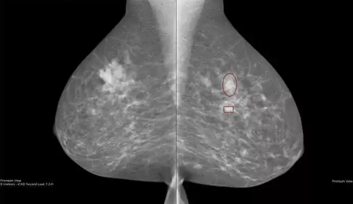
ProFound AI for 2D Mammography | Image Source
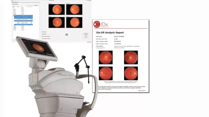
3. IDx-DR
IDx, the maker of autonomous AI diagnostic tools, built IDx-DR. It is a device capable of diagnosing diabetic retinopathy without human intervention.
The system is the first FDA-approved autonomous artificial intelligence (AI) that uses software to analyze images from a retinal camera called the Topcon NW400 for evidence of lesions.
How does it work?
A physician uploads the digital images of the patient’s retinas to a cloud server on which IDx-DR software is installed. The software can offer accurate results for further diagnostic evaluation.
The FDA designated IDx-DR as a Breakthrough Device. In this context, Michael Abramoff, the company’s founder and president, aptly stated that the healthcare system needed more efficient and cost-effective ways to detect diabetic retinopathy.
4. OsteoDetect (Imagen Technologies)
Imagen Technologies’ OsteoDetect software is a computer-aided detection and diagnostic software. The technology uses an artificial intelligence algorithm to examine and analyze 2D X-ray images for any indications of distal radius fracture.
The software uses deep learning techniques to analyze wrist radiographs (post-anterior [PA] and lateral [LAT]) for distal radius fractures in adult patients. Furthermore, it marks the location of the fracture on the image to aid the provider in detection and diagnosis.
The FDA-approved OsteoDetect is intended to be used by clinicians in various settings, including primary care, urgent care, emergency medicine, and specialty care, such as orthopedics.
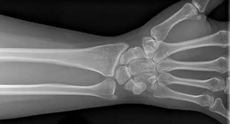
OsteoDetect AI-Guided Software | Image Source
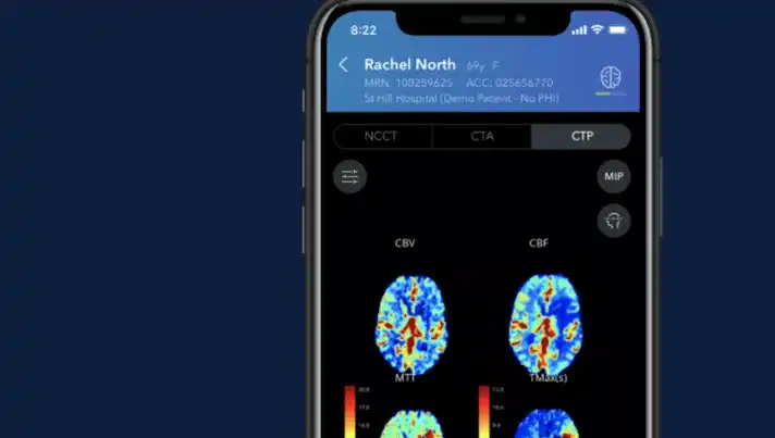
5. ContaCT (Viz.ai)
Viz.ai’s ContaCT is a notification-only, parallel workflow tool. ContaCT uses an artificial intelligence algorithm to examine and analyze computed tomography (CT) images for biomarkers symptomatic of a possible stroke.
The software uses deep learning to quantify image characteristics and perform vessel segmentation that is consistent with large vessel occlusions (LVOs) in scans.
Time is critical when treating strokes, so a mobile app that sends notifications directly to the treating physician can be life-saving.
Can a Shortage of Trained and Experienced Radiologists Be Solved with AI?
On the one hand, there is an increase in demand for cross-sectional imaging (CT and MRI). On the other, there is a lack of trained radiologists to examine and analyze the images.
Across Europe, there is a massive scarcity of trained radiologists. The UK is facing the worst capacity constraints, with the lowest number of practicing radiologists per capita.
Budgetary constraints and an aging population coupled with the time-consuming process of image analysis are probably the reasons for the catastrophic shortage of radiologists across Europe. (Source)
With AI and machine learning, experts hope to address this problem. AI can be used to automate several manual activities, such as recording and running analysis over the data. However, there are still some challenges for using AI-based medical imaging software.
Challenges of Using AI in Medical Imaging
Global AI-enabled imaging modalities are poised to change the market landscape. However, AI-based medical imaging software faces some significant challenges.
Let’s check the facets of crisis.
- Regulatory Challenges
AI applications need regulations for safety, privacy protection, and ethical use of sensitive information.
The intent behind AI’s design needs consideration, as some devices can be programmed to perform in unethical ways. So, regulations need to be set in a timely and relevant manner.
- A rise in Medical Imaging Expenses
The global market of artificial intelligence in medical imaging is expected to reach a projected value of USD 264.85 billion by 2026. According to research, the high cost and reluctance of adopting these systems are also expected to restrain the market growth. (Source)
The challenges also include new technology disrupting the provision of care and the need to improve patient outcomes. However, with the growing technological complexities and challenges, there is an excellent opportunity to harness technology and offer solutions to bridge the imaging capacity gap. As appreneurs, one can evolve by imbibing best practices to meet the growing needs.
Wrapping Up
Without a doubt, AI for medical imaging is at an exciting crossroads. AI is boosting the power of processing a massive number of medical images and has a promising future. Despite the excitement, enhancements are needed before it becomes more robust. Yet, AI can play a significant role in the medical imaging space. It can change the way people process the enormous number of images, improving patient care and reducing scan times.
We’re still scratching the surface with respect to AI’s capabilities. The medical imaging landscape will accelerate with growing customer confidence in AI-based clinical solutions. All you need to do is keep developing robust software that can enable radiologists to increase diagnostic accuracy.
Build a Blockbuster AI-enabled Medical Imaging Software with Imaginovation
Despite the many challenges, it is exciting to bring medical imaging AI solutions to the market. It is evident that radiology AI will grow in leaps-and-bounds. So, if you are looking at developing robust medical imaging software, talk to us.
We are an award-winning web and mobile app development company in Raleigh, with an incredible experience of developing sustainable and game-changing digital stories. Let’s talk.


